- Tour Home
- >
- Equine Hospital
- >
- Diagnostic Imaging
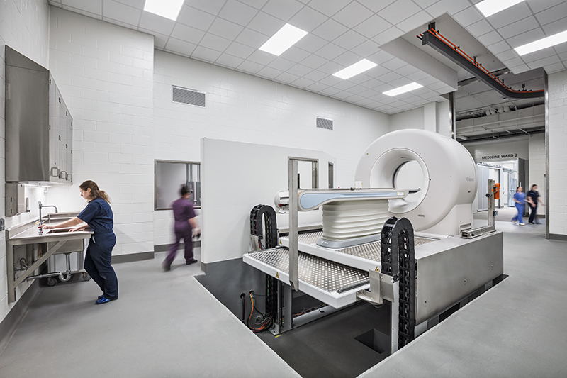
Diagnostic Imaging
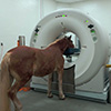
The equine diagnostic imaging center which consists of separate rooms for Computed Tomography (CT), Magnetic Resonance Imaging (MRI), Radiology and Nuclear Imaging. We are fortunate to have imported from Germany the Qalibra standing CT unit. This unit provides high-quality CT imaging without the need for general anesthesia. There are only three of these units in the United States and Purdue has two of them – one here and one at our Caesars Entertainment Equine Specialty Hospital in Shelbyville, Indiana.
The MRI unit is a 1.5 Tesla machine and functions by producing a powerful magnetic field that allows for the visualization of various body tissues. MRI is used to diagnose medical problems just as in human medicine.
Nuclear imaging tags specific sites (bone or glandular tissue) in the body and allows the clinician to visualize the area.
Ultrasound is performed in an exam room by one of the clinicians. All radiographs, ultrasounds, CTs and MRIs can be viewed on any computer in the hospital and shown to clients or utilized for teaching purposes.